Spray Smart, Seal Fast: A Practical Playbook for Hemostatic Powder Application in OR, Endoscopy, and Prehospital Settings
Endoscopic hemostatic powders now achieve high immediate hemostasis rates in non‑variceal gastrointestinal bleeding, making them a go‑to bridge or adjunct when visualization is limited or bleeding is diffuse. In parallel, absorbable microporous polysaccharide hemostats (MPH) such as Arista AH are routine surgical adjuncts, prized for fast, incremental dosing under direct vision. Yet the same properties that make powders fast and flexible—fine particles, rapid wicking, broad plumes—also introduce pitfalls: caking from humidity, off‑target aerosol, and washout in torrential hemorrhage [5–7,13,22].
This article is a practical guide to spraying smarter and sealing faster. It provides stepwise readiness checks, applicator selection cues, plume‑control tactics, dosing choreography, and post‑use protocols tailored to the operating room (OR), GI endoscopy suite, and the prehospital setting—aligned with device IFUs and field guidelines. You’ll learn how to set up and stage powders to avoid flow variability, match dose to bleeding rate, control aerosol spread, manage high‑flow scenarios without intravascular risk, and integrate monitoring informed by MAUDE signals.
Architecture/Implementation Details
Pre‑use readiness: humidity, desiccants, integrity, sterile field
- Stage powders late and dry. Hygroscopic formulations cake in humid rooms, degrading flow and dose uniformity; keep containers sealed until use and maintain desiccant‑protected packaging intact [5,6,13].
- Verify integrity: check pouches for seal breaches or moisture fogging, confirm sterility indicators, and inspect applicators for cracks or clogged nozzles. Discard compromised units per IFU [5–7].
- Prepare a sterile landing zone. In the OR, set a dry suction field with low‑flow suction poised to capture off‑target plume without collapsing the nascent clot. In endoscopy, ensure a dry catheter and purge lines before entering the patient [6,7].
- Moisture and anti‑static basics: wipe external nozzles dry, avoid saline splash on tips, and ground the patient bed/OR table as available to reduce static‑induced bridging in powder paths [5,6,13].
Applicator selection and setup: bellows, cartridge, catheter
- Bellows applicators (OR) deliver short bursts with tactile feedback for incremental dosing under visualization. They pair well with MPH powders like Arista AH; nozzle options and simple diffusers help expand the footprint while lowering jet momentum.
- Cartridge/gas‑assisted systems (endoscopy) use CO2 and dedicated catheters to distribute powder broadly without clogging, critical for diffuse mucosal bleeding. EndoClot PHS and TC‑325 (Hemospray) provide controlled, wide coverage through catheterized delivery [6,7].
- Nozzle geometry matters: wider, diffuser‑style tips spread dose gently across irregular beds, while narrower tips target confined recesses. Avoid high‑momentum jets that can dislodge early clot [5–7].
- Setup checks: confirm CO2 cartridge pierce integrity, catheter patency, and that anti‑reflux valves function. Prime per IFU to avoid bolus surges at the first squeeze [6,7].
Plume control and protection: diffuser technique, shielding, suction
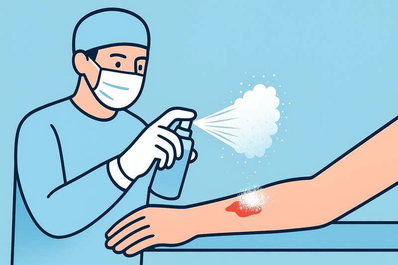
- Aim across, not into, the bleed. Start several centimeters back, using a sweeping, diffuse pass to create a dryable platform, then step closer for spot reinforcement. This reduces back‑spray and inhalable aerosol [6,7,13].
- Shield and suction smartly. Use clear eye protection and masks; coordinate low‑to‑moderate suction at plume edges to capture stray particles without stripping the forming layer [5–7,22].
- Restrict room turbulence. Pause adjacent irrigation and minimize airflow over the wound during application to limit off‑target aerosol dispersion, a key safety consideration given respirable fractions in fine powders [13,22].
Dosing and compression choreography: match mass to bleeding rate
- Work in increments. Lay light layers with continuous visualization; reassess after each burst. IFUs emphasize avoiding intravascular injection and clearing loose excess in confined spaces to reduce mass effect [5–7].
- Match dose to bleed. Faster rates need larger early mass to overcome flow and establish a barrier. For accessible external wounds, manual compression synergizes with powder mechanisms and remains central to hemostasis [1,5,9,10].
- Let the plug set. Once oozing slows, avoid high‑pressure irrigation. A gentle saline mist or passive suction at the periphery can help consolidate without lifting the layer [5–7].
Managing high‑flow hemorrhage: prevent washout, know when not to spray
- In torrential arterial bleeding, free powder alone is prone to washout before a gel matrix stabilizes. Prioritize mechanical control: direct pressure, tourniquets when indicated, and deep packing with hemostatic gauze; powders can serve as adjuncts only once flow is tamed [1,2,9,10].
- The Committee on TCCC specifically favors hemostatic gauze (kaolin or chitosan) over loose granular powders for field wound packing, citing depth deposition, compression synergy, and safety [1,2].
- Chitosan and kaolin dressings perform reliably under compression in swine extremity models, including during hypothermia and hemodilution—reinforcing the primacy of packing plus pressure in high‑flow scenarios [9,10,20].
Depth and geometry tactics: tracts, cavities, irregular beds
- Narrow tracts: avoid “dusting” the entrance. Use depth‑targeting tips when available; otherwise, prioritize gauze packing to deliver hemostatic material where the vessel bleeds [1,2].
- Cavities/irregular surfaces: pick a diffuser nozzle to blanket recesses. Begin with a broader first pass to create a drying substrate, then “spot‑spray” persistent points while limiting jet momentum [5–7].
- Endoscopic contours: catheterized systems excel in diffuse mucosal oozing across folds; maintain a few centimeters’ standoff to avoid mucosal disruption and maximize even coverage [6,7,21].
Residue and removal: intravascular avoidance, mass‑effect caution, documentation
- Never inject into vessels. Maintain visual confirmation of extravascular deposition and avoid direct cannulation of pulsatile sources with a narrow jet [5–7].
- Remove loose excess where mass effect matters (e.g., tight cranial, spinal, or orbital spaces). Absorbable MPH like Arista AH can be left in situ per IFU, but free loose piles should be reduced once hemostasis is achieved.
- Document location and estimated amount. Most powders are radiolucent; charting helps downstream teams interpret imaging and plan re‑entry if needed [5–7].
Comparison Tables
Applicator and delivery options
| Delivery mode | Typical setting | Coverage style | Strengths | Limitations | Representative devices |
|---|---|---|---|---|---|
| Bellows handheld | OR (open surgery) | Short, incremental bursts via nozzle/diffuser | Tactile dosing; simple; compatible with absorbable MPH | Sensitive to humidity; plume control requires suction | Arista AH applicators |
| Cartridge/gas‑assisted | GI endoscopy | Broad, CO2‑driven spray via catheter | Wide, even coverage; good for diffuse lesions | Requires catheter priming; gas management; clog risk if moist | EndoClot PHS; Hemospray TC‑325 [6,7] |
| Nozzle with depth tip | OR, select wounds | Directed placement into recesses | Improves deposition in irregular cavities | Limited availability; still not a substitute for packing | Available on some OR kits |
Powder classes commonly used in these workflows
| Class | Dominant mechanism | Best fit | Key cautions |
|---|---|---|---|
| MPH (starch) | Rapid dehydration; matrix swelling | OR adjunct under direct vision; endoscopic variants for mucosa | Washout in torrential bleeding without compression; remove loose excess in tight spaces [5–7,13] |
| Chitosan (powder/gauze) | RBC/platelet agglutination; wet adhesion | Field packing as gauze; adjunct where cascade is impaired | Loose granules not TCCC‑preferred for deep/junctional wounds; risk if intravascular [1,2,9,10] |
| Mineral TC‑325 (Hemospray) | Physical barrier, adsorption (non‑biopolymer) | Endoscopic bridge/adjunct for non‑variceal GI bleeding | Catheter priming; avoid intraluminal occlusion of ducts; follow IFU [7,21] |
Best Practices
OR workflow: adjunct spraying that respects the clot
- Pre‑brief on target anatomy and mass‑effect risks; stage low‑flow suction and eye/airway protection.
- Keep the field as dry as feasible, then apply light, sweeping passes with a diffuser tip, reassessing after each burst; avoid direct, high‑momentum jets at pulsatile points.
- Use manual compression where accessible to accelerate sealing; re‑apply in increments only if active oozing persists [1,5].
- Once hemostasis is achieved, gently clear loose excess in confined spaces and document location/amount; do not irrigate aggressively.
Endoscopic spray sequence for diffuse mucosal lesions
- Confirm catheter patency and prime per IFU; maintain a few centimeters’ standoff to create even coverage without mucosal trauma [6,7].
- Start with a broad pass to stabilize the surface, then target residual bleeders with short bursts. Coordinate low suction to skim off airborne plume, not the applied layer [6,7,21].
- Reassess at intervals; many cases achieve immediate hemostasis but may require adjunct therapy if rebleeding occurs.
Prehospital: TCCC‑consistent field use
- For junctional and extremity hemorrhage, pack deep with hemostatic gauze (kaolin or chitosan) and hold firm pressure; tourniquet when indicated. Loose free powders are generally not first‑line for deep wounds in combat care [1,2].
- If a superficial external bleed is selected for a powder adjunct, shield airways/eyes, apply light layers, and secure with pressure dressing. Avoid intravascular deposition and document use for receiving teams [1,2].
- Prioritize rapid evacuation and continuous reassessment; powders are adjuncts, not substitutes for definitive control.
Post‑application monitoring and safety
- Perform timed rebleed checks (e.g., 2–5 minutes, then at closure or pre‑transport), and reassess hemodynamics (HR, BP, MAP) to confirm systemic stabilization.
- Log device lot numbers and sites of application. Monitor for adverse events (airway exposure, embolic concerns) and feed back into training and IFU adherence; the FDA MAUDE database is a useful signal source for rare events and updates.
- In multi‑session care (e.g., staged endoscopy), note that many powders are radiolucent; charting the location and estimated mass aids downstream interpretation [5–7].
Practical Examples
- OR capillary oozing (hepatic bed):
- Dry the surface with gentle suction and sponges; pause irrigation.
- Open an MPH unit (e.g., Arista AH) at the field only when ready; confirm nozzle patency and attach a diffuser.
- From several centimeters away, deliver a light, sweeping pass to create a dehydrated layer. Reassess. If oozing persists, add short bursts to hot spots while keeping suction peripheral.
- Apply manual compression for 30–60 seconds if accessible. Once dry, gently remove loose piles in confined recesses. Document application, especially near critical structures [1,5].
- Endoscopic non‑variceal diffuse oozing (gastric body):
- Prime the CO2‑assisted powder system (EndoClot PHS or TC‑325) per IFU to avoid bolus surges [6,7].
- Position the catheter a few centimeters from the mucosa; perform a broad pass to stabilize the surface, then spot‑apply to residual oozers.
- Coordinate low suction to capture airborne plume without lifting the layer. Reassess at 1–2 minutes; many cases show immediate hemostasis, with re‑application as needed [6,7,21].
- Prehospital scalp laceration with brisk venous bleeding:
- Apply direct pressure and consider a pressure dressing. If using hemostatic adjuncts, pack with kaolin or chitosan gauze to the depth of the wound and compress firmly [1,2].
- Reserve loose powder for superficial surfaces only; shield the airway/eyes, apply minimal layers, compress, and secure. Continuous reassessment and rapid transport remain priorities. 🚑
Conclusion
Sprayable hemostatic powders are powerful adjuncts when deployed with discipline: dry‑chain storage, the right applicator for the anatomy, careful plume control, incremental dosing under visualization, and a bias toward packing and pressure when flow is torrential. OR teams can exploit absorbable MPH for fast, conformal control of capillary and venous oozing, while endoscopists benefit from catheterized, gas‑assisted coverage for diffuse mucosal lesions. In the field, TCCC‑consistent workflows keep hemostatic gauze at the center and reserve free powders for select superficial use. Across settings, avoid intravascular deposition, clear loose excess where mass effect matters, document radiolucent residues, and keep training current with MAUDE signals.
Key takeaways:
- Keep powders dry until use; stage suction and PPE to manage plume [5–7,13,22].
- Choose bellows for OR precision and catheterized systems for diffuse endoscopic coverage [5–7].
- Dose incrementally and pair with compression; for high‑flow bleeds, prioritize packing and pressure per TCCC [1,2,9,10].
- Avoid intravascular application; remove loose excess in tight spaces and document radiolucent residues [5–7].
- Monitor for rebleed and adverse events; feed observations back into training and IFUs.
Next steps: standardize team checklists for pre‑use readiness, create setting‑specific spray protocols, and integrate adverse‑event reviews into quarterly training. Looking ahead, expect diffuser and catheter refinements that further broaden coverage while reducing jet momentum—and clearer, evidence‑based guidance on plume metrics and depth deposition as studies evolve.
Sources
- Deployed Medicine – Hemorrhage Control (TCCC): https://deployedmedicine.com/market/11/content/53 — Field guidelines that prioritize hemostatic gauze and compression over loose granular powders in deep/junctional hemorrhage.
- Joint Trauma System – Committee on TCCC (CoTCCC): https://jts.health.mil/index.cfm/committees/cotccc — Official TCCC recommendations and adjuncts for prehospital hemorrhage control that inform workflow and device selection.
- Baxter – Arista AH (Microporous Polysaccharide Hemostat): https://advancedsurgery.baxter.com/arista-ah — OR IFU cues on absorbable MPH use, dosing, removal of excess, and storage/desiccant practices.
- EndoClot Plus – Polysaccharide Hemostatic System (PHS): https://endoclot.com/products/phs/ — Endoscopic, CO2‑assisted, catheterized powder delivery details that underpin setup, priming, and coverage strategies.
- Cook Medical – Hemospray (TC‑325) Endoscopic Hemostatic Powder: https://www.cookmedical.com/products/hemospray/ — Endoscopic spray workflow and plume considerations for diffuse mucosal bleeding control.
- Kheirabadi et al., 2011 (J Trauma): https://pubmed.ncbi.nlm.nih.gov/21795879/ — Preclinical extremity arterial hemorrhage data supporting the efficacy of hemostatic dressings under compression.
- Kheirabadi et al., 2009 (J Trauma): https://pubmed.ncbi.nlm.nih.gov/19854364/ — Evidence for hemostatic dressing performance during hypothermia and hemodilution, reinforcing compression‑first in high‑flow states.
- Recent Advances in Hemostatic Materials for Wound Healing (Bioactive Materials, 2021): https://www.sciencedirect.com/science/article/pii/S2452199X20303639 — Mechanistic context (dehydration, adhesion), humidity/caking risk, and aerosolization considerations across powder classes.
- Kaolin/Contact Pathway Activation Background: https://pubmed.ncbi.nlm.nih.gov/15273965/ — Mechanistic rationale for kaolin gauze in field packing and compression workflows.
- EndoClot PHS Clinical Experience (PubMed): https://pubmed.ncbi.nlm.nih.gov/34224833/ — Clinical evidence for high immediate hemostasis rates with catheterized powders in non‑variceal GI bleeding.
- FDA MAUDE Adverse Event Database: https://www.accessdata.fda.gov/scripts/cdrh/cfdocs/cfmaude/search.cfm — Post‑market signals used to inform plume protection, airway safety, and training updates.