From DDA to FXII Activation: The Architecture and Performance Physics of Spray‑On Biopolymer Hemostats
A swine extremity hemorrhage benchmark still frames this field: modern chitosan agents and starch‑based microporous spheres regularly achieve rapid hemostasis when applied correctly, even under shock‑mimicking derangements. Meanwhile, endoscopic powders deliver through CO2‑assisted catheters to coat bleeding mucosa without clogging, highlighting how delivery physics can be as determinative as chemistry. As Tactical Combat Casualty Care (TCCC) continues to prioritize hemostatic gauze for deep packing, sprayable biopolymer powders have sharpened their edge in open surgical and luminal settings by optimizing particles, charge, and plume geometry.
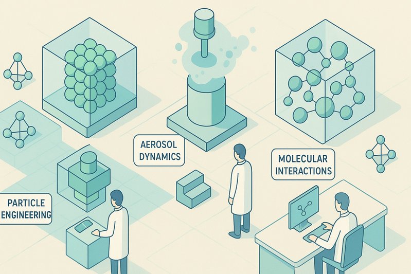
This article maps the engineering stack—from polymer family mechanisms to particle microstructure, interfacial adhesion chemistries, contact pathway accelerants, and aerosol dynamics—that governs the performance of spray‑on biopolymer hemostats. We will show how tuning degree of deacetylation (DDA), porosity, crosslinking, and nozzle design translates into measurable clot metrics such as early mechanical sealing and TEG/ROTEM signatures. Readers will learn where each class excels, how device physics shapes deposition in complex cavities, and how to interpret functional outputs against challenging physiologies.
Architecture/Implementation Details
Mechanistic architecture by class
- Chitosan agglutination. Cationic amines electrostatically bind negatively charged RBCs and platelets, yielding a rapid, cascade‑independent primary plug and strong mucoadhesion on wet tissue. Charge density rises with DDA, increasing agglutination potency but imposing a cytocompatibility trade‑off; molecular weight (MW) influences the viscosity and film strength of the resulting wet gel. Modern chitosan powders also leverage intrinsic antimicrobial activity at the tissue interface.
- MPH (microporous polysaccharide spheres) dehydration. Engineered, highly porous starch spheres wick plasma and water, locally concentrating platelets and clotting proteins while swelling into a conformal mechanical matrix. The mechanism is largely physical—fast capillary uptake and sequestration—making MPH highly effective under direct visualization and incremental dosing paradigms.
- Alginate gelation. Calcium‑alginate gels rapidly on contact, simultaneously delivering Ca2+ to support coagulation and forming a conformal hydrogel that resists washout when supported by adhesion partners (e.g., chitosan).
- Collagen/gelatin platelet cues. Collagen‑derived scaffolds present platelet activation ligands (e.g., GPVI binding), accelerating platelet activation and aggregation; gelatin microsponges milled to powders add porosity and capillary pathways for early seal formation.
- Fibrin end‑stage formation. Co‑delivered thrombin/fibrinogen within composite particulates can drive direct fibrin polymerization at the wound, bypassing upstream deficits. While most clinical fibrin products are liquid sprays, particulate co‑delivery shows promise for rapid, terminal clot formation.
Microstructure as a performance lever
Particle size distribution and porosity/aerogel‑like morphologies set wicking kinetics, adsorption surface area, and the speed/strength of the initial mechanical seal. Highly porous microspheres dramatically shorten the path length for capillary flow and protein adsorption, while microfiber chitosan architectures bridge irregular topographies to form cohesive wet gels. Ultra‑fine fractions boost surface area but raise respirable particle risk, shaping requirements for diffuser nozzles and plume management to protect airways and eyes.
Early mechanical sealing depends on three coupled processes: (1) fluid sequestration to concentrate clotting factors (MPH/alginate/cellulose pathways), (2) cellular agglutination and adhesion (chitosan/catechol/TA), and (3) nascent fibrin formation if contact activation (kaolin/nanosilicates) or exogenous thrombin are present. Managing particle architectures to execute at least two of these in the first seconds is the hallmark of best‑in‑class powders.
Charge engineering in chitosan
- DDA and charge density. Higher DDA increases cationic sites and RBC/platelet binding, expediting plug formation when enzymatic coagulation is impaired (hypothermia, acidosis, anticoagulation). Excessive charge density, however, risks local cytotoxicity—necessitating a cytocompatibility window tuned by DDA, MW, and blending with polyanions (e.g., alginate).
- MW and wet‑gel film strength. Higher MW enhances chain entanglement and viscoelasticity of the post‑wetting gel, improving shear resistance under flow. Sterilization choices matter: gamma irradiation can depolymerize chitosan, lowering MW and viscosity, potentially diminishing cohesive strength and adhesion; EtO avoids scission but requires rigorous aeration to remove residues.
Crosslinking and interfacial adhesion under flow
- Calcium‑alginate gelation drives fast ionic crosslinking with simultaneous Ca2+ provision to the cascade.
- Catechol/tannic acid (TA) reinforcement furnishes underwater adhesion via covalent, hydrogen‑bonding, and metal‑coordination interactions with tissue and proteins; TA also complexes proteins and can aggregate cells, boosting early plug stability and contamination tolerance.
- Polyelectrolyte complexes (PECs) such as chitosan–alginate or chitosan–oxidized cellulose tune local pH, swelling, and cohesion, creating composite matrices that resist shear and reduce rebleed on irregular wound beds.
Contact pathway accelerants and adsorption physics
- Kaolin and synthetic nanosilicates accelerate intrinsic pathway activation by providing negatively charged, high‑surface‑area templates that trigger factor XII (FXII) activation, shortening clot initiation (e.g., TEG/ROTEM R time) and supporting a denser fibrin network. Particle safety becomes central here—engineers must cap aerodynamic diameters and control fines to mitigate inhalation/embolization risks while maintaining adsorption efficacy.
Aerosol delivery physics and deposition profiles
Plume dynamics set where particles land and how strongly they impact. Key variables include nozzle diameter, diffuser geometry, standoff distance, pulse duration, and whether delivery is CO2‑assisted.
- OR bellows/cartridge applicators allow incremental dosing under visualization; broad diffusers reduce jet momentum to avoid dislodging nascent gel.
- CO2‑assisted catheters (e.g., EndoClot PHS) deliver powders through long, hydrophobic lumens into GI lumens with wide, gentle plumes to coat diffuse bleeding surfaces without clogging—an instructive model for cavity coverage.
- Plume control balances cavity penetration with minimization of airborne dispersion; anti‑static paths and flow aids reduce bridging, while desiccant packaging preserves free‑flow characteristics.
Dose dynamics and reapplication logic
Hemostatic mass must exceed local bleed rate and resist washout until biological stabilization. Best performance comes from:
- Sufficient initial mass to create a continuous matrix across the bleed source(s), with prompt reapplication if focal oozing persists.
- Compression and/or packing when feasible—especially in high‑flow spurting—because even excellent powders can be physically displaced before gel networks or agglutinative films mature.
- Avoidance of intravascular injection and removal of excess material in confined spaces to limit mass effect, per IFUs.
Functional readouts and cross‑material harmonization
- TEG/ROTEM: Contact activators (kaolin/nanosilicates) primarily reduce R time (clot initiation), occasionally increasing alpha angle and MA via more rapid thrombin generation; chitosan’s early sealing may be under‑represented in these viscoelastic assays yet manifests clinically as reduced rebleed despite minimal change in enzymatic kinetics. Harmonized protocols and composite endpoints that couple viscoelastic metrics with early mechanical seal assessments would better compare across material classes.
Comparative performance envelopes under physiologic stress
- Hypothermia/acidosis/anticoagulation: Chitosan’s cascade‑independent cellular agglutination and mucoadhesion maintain efficacy where enzymatic steps falter; kaolin gauze remains effective under compression but may exhibit slowed initiation in severe acidosis.
- Arterial spurting: MPH excels in controlled fields but benefits from compression or packing to prevent washout before a matrix stabilizes.
- Contaminated, constantly wet tissue: Chitosan and catechol/TA‑functionalized composites offer stronger wet adhesion and potential antimicrobial advantages, aiding residence time and early plug stability.
🔬 Diagram concept (textual): A split schematic shows (left) porous microsphere packing that wicks plasma and concentrates cells/proteins, and (right) cationic chitosan chains bridging RBCs/platelets while catechol anchors to tissue proteins; an overlay plume map depicts diffuser‑widened, low‑momentum deposition versus narrow high‑momentum jets.
Comparison Tables
Mechanisms by class and expected early behavior
| Class | Dominant early mechanism | Cascade dependence | Early strengths | Potential limitations |
|---|---|---|---|---|
| Chitosan powders | Electrostatic RBC/platelet agglutination; mucoadhesion | Low (works despite coagulopathy) | Fast primary plug; adheres to wet tissue; antimicrobial | Charge‑toxicity trade‑off; embolization risk if intravascular |
| MPH (starch) | Rapid dehydration; protein/platelet concentration; matrix swelling | Indirect (concentration‑driven) | Quick coverage; absorbable; OR workflow | Washout in torrential flow without compression |
| Alginate (Ca‑alginate) | Ionic gelation; Ca2+ provision | Moderate (supports cascade) | Conformal gels; pairable with chitosan adhesion | Needs adhesion partner for shear resistance |
| Collagen/gelatin | Platelet activation/scaffold (GPVI cues) | Moderate | Strong platelet engagement; porous architecture | Immunogenicity and swelling considerations |
| Kaolin/nanosilicate additives | FXII contact activation; adsorption | High (intrinsic pathway) | Shorter R time; denser fibrin | Inhalation/embolization control required |
Aerosol/delivery variables and their effects
| Variable | Increase leads to | Hemostatic impact | Risk/constraint |
|---|---|---|---|
| Nozzle diameter | Higher mass flow, larger droplets | Faster coverage of large areas | Potential to dislodge nascent gel if jet momentum rises |
| Diffuser area | Broader, lower‑momentum plume | Gentle coating of complex cavities | Reduced penetration depth if over‑diffused |
| CO2 pressure | Longer throw, improved catheter transit | Better reach in lumens; stable powder flow | Airway dispersion risk; require pulse control |
| Particle porosity | Faster capillary wicking | Shorter seal initiation | Increased respirable fraction; packaging sensitivity |
Best Practices
- Engineer for dual‑mode initiation. Combine a fast physical pathway (dehydration or gelation) with adhesive or contact activation to stabilize the plug before shear dislodges it.
- Tune chitosan DDA and MW for a cytocompatible, high‑adhesion window; verify sterilization effects—avoid over‑irradiation that reduces MW and wet‑gel strength.
- Use polyelectrolyte complexes (e.g., chitosan–alginate) or catechol/TA functionalization to upgrade wet adhesion and cohesion in shear, particularly on contaminated or constantly wet surfaces.
- Control particle size distribution to maximize surface area and wicking while limiting respirable fines; pair with diffuser nozzles and CO2 pressure modulation to minimize jet momentum and off‑target aerosol.
- Align dose and reapplication with bleed rate and anatomy; in high‑flow spurting, combine with compression/packing when feasible and follow IFUs to avoid intravascular delivery.
- Interpret TEG/ROTEM in context: expect R‑time shortening with contact activators; for chitosan, track early mechanical sealing and rebleed rates alongside viscoelastic metrics.
Practical Examples
1) Quick dose calculus for initial application (conceptual)
# Hemostat dose estimator (illustrative only)
# Inputs: bleed_rate (mL/min), target_seal_time (min), powder_uptake_ratio (mL blood bound per g powder)
# Output: minimum powder mass (g) for initial barrier formation

def estimate_mass(bleed_rate, target_seal_time, uptake_ratio):
 blood_to_sequester = bleed_rate * target_seal_time # mL
 safety_factor = 1.5 # to cover heterogeneity and shear
 return safety_factor * (blood_to_sequester / uptake_ratio)

# Example: 80 mL/min spurting bleed, aim to stabilize in 1.0 min,
# powder binds 10 mL/g (porous starch or chitosan composite)
print(round(estimate_mass(80, 1.0, 10.0), 1), 'g') # -> 12.0 g
Rationale: dehydrating/adsorbing powders must sequester sufficient fluid to bridge to biological stabilization; agglutinative powders benefit from mass that ensures contiguous coverage and bridging across the bleeding source. Always reassess after the first pulse and reapply incrementally as needed.
2) Applicator setup sketch for gentle, wide‑area coating
applicator:
 mode: CO2_assisted # catheter delivery in luminal/cavity bleeds
 co2_pressure: 7-10 psi # moderate to avoid high jet momentum
 nozzle:
 type: radial_diffuser
 outlet_id: 3.0 mm # wider for low-velocity plume
 pulse:
 duration_ms: 300-500 # short bursts to layer powder
 interval_ms: 800-1200 # allow wetting/consolidation between pulses
 standoff_distance: 10-30 mm # under visualization when possible
 anti_static_path: enabled
 desiccant_packaging: verified_intact
Principle: broaden the plume and lower momentum to avoid dislodging early gels/aggregates, especially with highly porous powders. Use short pulses to let capillary wetting and adhesion work before reapplication; maintain moisture‑protected flow for dose uniformity. ⚙️
Conclusion
Spray‑on biopolymer hemostats succeed by stacking mechanisms: rapid fluid sequestration and ionic gelation, cellular agglutination and wet adhesion, and contact‑pathway acceleration where safe and appropriate. Microstructure and charge design determine how quickly an early mechanical seal forms; crosslinking and catechol/TA chemistries maintain cohesion under flow; and aerosol physics decides whether the material lands where it matters. In hypothermia, acidosis, or anticoagulation, chitosan’s cascade‑independent action provides resilience; MPH and alginate excel under visualization with disciplined dosing and plume control. Interpreting TEG/ROTEM alongside early mechanical sealing yields a truer cross‑material comparison.
Key takeaways:
- Optimize particles (size/porosity) and plume (diffuser/pressure) together—they’re co‑determinants of early sealing.
- Balance chitosan DDA/MW for adhesion without cytotoxicity; verify sterilization impacts on chain length and gel strength.
- Pair dehydration/gelation with adhesion or contact activation to resist washout in high‑shear bleeding.
- Read TEG/ROTEM in mechanism‑aware fashion; add mechanical seal metrics for fairness across classes.
Next steps for practitioners and developers:
- Validate aerosol plumes with respirable‑fraction and deposition tests; tune diffuser and pulse recipes.
- Build PEC/catechol‑reinforced composites that combine fast uptake with robust wet adhesion.
- Incorporate safety‑minded contact activators and rigorously cap fines; align ISO 10993 programs accordingly.
Looking ahead, multi‑mechanism composites—catechol‑reinforced chitosan/alginate with carefully controlled nanosilicate contact activators—paired with adjustable CO2 diffusers promise faster initiation, stronger shear resistance, and safer, more predictable deposition into complex wound geometries.
Sources
- url: https://deployedmedicine.com/market/11/content/53 title: Deployed Medicine – Hemorrhage Control (TCCC) relevance: Establishes field best practices, compression/packing synergy, and cautions about loose powders—key for dose/reapplication logic and deployment context.
- url: https://jts.health.mil/index.cfm/committees/cotccc title: Joint Trauma System – Committee on TCCC (CoTCCC) relevance: Confirms guideline emphasis on hemostatic gauze and informs where sprayable powders fit relative to packing in external hemorrhage.
- url: https://www.teleflex.com/usa/en/product-areas/hemostasis/quikclot/index.html title: Teleflex QuikClot (Kaolin Hemostatic Gauze) relevance: Reference for kaolin contact activation and as a performance comparator under compression.
- url: https://www.celoxmedical.com/products/celox-granules/ title: Celox Medical – Celox Granules (Chitosan Hemostat) relevance: Illustrates chitosan agglutination mechanism and use in high‑bleed scenarios.
- url: https://advancedsurgery.baxter.com/arista-ah title: Baxter – Arista AH (Microporous Polysaccharide Hemostat) relevance: Supports MPH dehydration mechanism, porosity/wicking design, and OR applicator paradigms.
- url: https://endoclot.com/products/phs/ title: EndoClot Plus – Polysaccharide Hemostatic System (PHS) relevance: Documents CO2‑assisted catheter delivery, plume control, and broad mucosal coverage relevant to aerosol physics.
- url: https://pubmed.ncbi.nlm.nih.gov/21795879/ title: Kheirabadi et al. Comparison of Hemostatic Dressings in a Swine Model of Extremity Arterial Hemorrhage (J Trauma, 2011) relevance: Provides preclinical performance benchmarks and endpoints like time to hemostasis.
- url: https://pubmed.ncbi.nlm.nih.gov/19854364/ title: Kheirabadi et al. Efficacy of Topical Hemostatic Dressings in Hypothermia and Hemodilution (J Trauma, 2009) relevance: Demonstrates chitosan’s resilience under coagulopathic conditions, informing the comparative envelope.
- url: https://www.fda.gov/regulatory-information/search-fda-guidance-documents/use-international-standard-iso-10993-1-biological-evaluation-medical-devices-part-1 title: FDA Guidance – Use of ISO 10993‑1 for Biological Evaluation of Medical Devices relevance: Frames biocompatibility evaluation for high‑charge chitosan and nano‑enabled additives.
- url: https://www.mdpi.com/1422-0067/20/7/1693 title: Antimicrobial Activity of Chitosan and Its Derivatives (Int J Mol Sci, 2019) relevance: Supports chitosan’s cationic interactions, antimicrobial effects, and charge‑dependent cytocompatibility considerations.
- url: https://www.sciencedirect.com/science/article/pii/S2452199X20303639 title: Recent Advances in Hemostatic Materials for Wound Healing (Bioactive Materials Review, 2021) relevance: Comprehensive technical review of polymer classes, microstructure effects, crosslinking/adhesion chemistries, and device variables.
- url: https://onlinelibrary.wiley.com/doi/10.1002/adhm.202000711 title: Mussel‑Inspired Tissue Adhesives for Hemostasis (Adv Healthcare Mater, 2020) relevance: Details catechol/TA wet‑adhesion mechanisms and reinforcement strategies for powders.
- url: https://www.ncbi.nlm.nih.gov/pmc/articles/PMC4373602/ title: Thromboelastography and ROTEM: Methodology and Clinical Applications (Transfus Med Rev, 2014) relevance: Grounds interpretation of R time, alpha, and MA when comparing contact activators vs agglutinative materials.
- url: https://pubmed.ncbi.nlm.nih.gov/15273965/ title: Kaolin/Contact Pathway Activation Background relevance: Explains FXII activation on negatively charged surfaces, central to kaolin/nanosilicate additives.
- url: https://www.accessdata.fda.gov/scripts/cdrh/cfdocs/cfmaude/search.cfm title: FDA MAUDE Adverse Event Database relevance: Supports aerosol safety emphasis (inhalation/embolization vigilance) for sprayable powders.
- url: https://pubmed.ncbi.nlm.nih.gov/19021290/ title: Effects of Gamma Irradiation on Chitosan Molecular Weight/Properties relevance: Documents sterilization‑induced MW reduction and its implications for gel strength and adhesion.